Category:Histology
Jump to navigation
Jump to search

Histology is the study of the microscopic anatomy of cells and tissues of plants and animals. Place files relating to normal, disease-free tissues in a suitable subcategory of "Category:Animal histology", "Category:Human histology" or "Category:Plant histology".
study of the microscopic anatomy of cells and tissues of plants and animals  | |||
| Upload media | |||
| Instance of | science | ||
|---|---|---|---|
| Different from | |||
| |||
- Place files relating to diseased tissues in a suitable subcategory of "Category:Histopathology":
- Diseased tissues of human beings should be placed in a suitable subcategory of "Category:Human histopathology".
- Diseased tissues of animals should be placed in a suitable subcategory of "Category:Veterinary histopathology".
Subcategories
This category has the following 31 subcategories, out of 31 total.
*
.
?
A
B
C
D
F
G
H
I
M
P
S
T
Media in category "Histology"
The following 199 files are in this category, out of 199 total.
 201904 myotube.svg 512 × 410; 2.13 MB
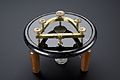
 Apparatus for preparing injected preparations Wellcome M0018212.jpg 2,717 × 3,889; 1.59 MB
 Box of 126 microscope preparations of a spinal column, Edinb Wellcome L0057901.jpg 2,832 × 4,256; 1.04 MB
_-_IHM-0651.jpg/120px-Boîte_de_coupes_histologiques_(physiologie)_-_IHM-0651.jpg) Boîte de coupes histologiques (physiologie) - IHM-0651.jpg 3,600 × 2,612; 2.09 MB
Boîte de coupes histologiques (physiologie) - IHM-0651.jpg 3,600 × 2,612; 2.09 MB
 Boîte de coupes histologiques - IHM-0254.jpg 3,088 × 2,056; 2.9 MB
 Brockhaus and Efron Encyclopedic Dictionary b65 328-1.jpg 3,135 × 2,625; 1.52 MB
 Brockhaus and Efron Encyclopedic Dictionary b65 328-2.jpg 3,403 × 2,774; 1.72 MB
 Brockhaus and Efron Encyclopedic Dictionary b65 328-3.jpg 2,953 × 2,563; 1.01 MB
 Brockhaus and Efron Encyclopedic Dictionary b65 328-4.jpg 3,554 × 2,803; 924 KB
 Brockhaus and Efron Encyclopedic Dictionary b65 328-5.jpg 1,594 × 2,277; 317 KB
 Cambium2.jpg 480 × 324; 29 KB
 Camilod facility.png 772 × 410; 623 KB
 Cellules, tissus, organes et systèmes.jpg 1,754 × 2,480; 842 KB
 Chromo.jpg 561 × 384; 65 KB
 Circumvallate.jpg 2,048 × 1,536; 595 KB
.jpg/120px-Cluster_(rus).jpg) Cluster (rus).jpg 1,296 × 433; 151 KB
Cluster (rus).jpg 1,296 × 433; 151 KB
.jpg/120px-Cluster_(ukr).jpg) Cluster (ukr).jpg 2,592 × 866; 399 KB
Cluster (ukr).jpg 2,592 × 866; 399 KB
 Comparison of cancer cell lines.png 640 × 285; 11 KB
 Compound monocular microscope, Vienna, Austria, 1831-1870 Wellcome L0057245.jpg 2,832 × 4,256; 1.28 MB
 Conger type callus 3ms White Light.TIF 2,048 × 1,536; 9.01 MB
 Coupe totale d'araignée.jpg 1,743 × 1,122; 217 KB
 Cresyl violet in weigh boat.jpg 2,994 × 2,349; 1,013 KB
 Cryostat Stage.jpg 1,632 × 1,224; 720 KB
 Cryptosporidium parvum auramine-rhodamine labeled.jpg 300 × 308; 6 KB
 Dense connective tissue-400x.jpg 2,048 × 1,536; 2.19 MB
 ElastinisJ.A.JPG 1,332 × 1,200; 393 KB
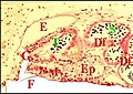
 Endocrinoide mâle 2.jpg 495 × 325; 32 KB
 Endoderm2 hr.png 400 × 290; 62 KB
 Endoderm2-ar.png 270 × 198; 30 KB
 Endoderm2.png 270 × 198; 37 KB
 Epidermis histology 2014.jpg 4,912 × 3,264; 710 KB
 ErbB2.jpg 2,048 × 1,536; 779 KB
- Es-Agruras-article.ogg 5 min 59 s; 4 MB
 FatStemCells.gif 260 × 208; 29 KB
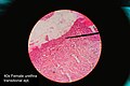 Female urethra histology.jpg 4,912 × 3,264; 994 KB
 Figures showing cell division. Wellcome M0016974.jpg 5,060 × 2,156; 2.73 MB
 Filistata insidiatrix, région épigastrique.jpg 3,581 × 2,525; 766 KB
 Freezing microtome, London, England, 1883-1885 Wellcome L0058209.jpg 4,256 × 2,832; 1.25 MB
 Frozen tissue array block.jpg 3,072 × 2,304; 1.04 MB
 Frozen tissue array section.jpg 1,077 × 680; 252 KB
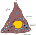 G cell miguelferig.png 1,404 × 1,393; 95 KB
 GAFA 1996-01-3-1 Str 51 Bone 200fach XPL.jpg 2,592 × 1,944; 1.15 MB
 Glande rostrale de Diguetia.jpg 4,171 × 3,427; 892 KB
 Glande venin Diguetia 1.jpg 3,525 × 2,430; 1.1 MB
 Glande venin Diguetia 2.jpg 1,836 × 1,237; 363 KB
 Glande venin Diguetia 3.jpg 1,867 × 1,179; 405 KB
 Glandes Scytodes 1.jpg 4,312 × 3,417; 815 KB
 Glandes Scytodes 2.jpg 5,029 × 3,370; 1.07 MB
_-_copia.jpg/107px-Glandula_sebasea_(3)_-_copia.jpg) Glandula sebasea (3) - copia.jpg 529 × 592; 203 KB
Glandula sebasea (3) - copia.jpg 529 × 592; 203 KB
 GlaudusisAudinys.JPG 1,600 × 1,200; 377 KB
 Globet cell miguelferig.png 921 × 1,395; 98 KB
 Globus pallidus and putamen - very low mag.jpg 4,272 × 2,848; 5.93 MB
 Glucagon rednblue.png 863 × 557; 1.29 MB
 Grasa parda vista al microscopio electrónico.tif 676 × 500; 1.11 MB
 Gray964-zh.png 402 × 600; 154 KB
 Haematoxylin powder.jpg 2,716 × 2,673; 1.28 MB
 Healthy mammary gland.jpg 250 × 186; 31 KB
 Hem.jpg 304 × 228; 42 KB
 Heme.jpg 304 × 228; 41 KB
 Hersilia sp., région épigastrique.jpg 3,165 × 2,074; 656 KB
 Hiperplasia wiki.PNG 644 × 234; 358 KB
 Histol.Technik.jpg 3,636 × 1,364; 1.01 MB
 Histologic Slide under MIcroscope.jpg 2,848 × 4,288; 2.84 MB
 Histological Architecture of the Joint Surface.jpg 576 × 359; 34 KB
.jpg/120px-Histologie_(1).jpg) Histologie (1).jpg 2,140 × 1,027; 1.01 MB
Histologie (1).jpg 2,140 × 1,027; 1.01 MB
.jpg/120px-Histologie_(3).jpg) Histologie (3).jpg 4,937 × 2,250; 1.51 MB
Histologie (3).jpg 4,937 × 2,250; 1.51 MB
.jpg/120px-Histologie_Basalmembran_(2).jpg) Histologie Basalmembran (2).jpg 2,149 × 1,226; 679 KB
Histologie Basalmembran (2).jpg 2,149 × 1,226; 679 KB
 Histologie Basalmembran.jpg 2,149 × 1,226; 674 KB
.jpg/120px-Histology_(1).jpg) Histology (1).jpg 2,700 × 1,800; 966 KB
Histology (1).jpg 2,700 × 1,800; 966 KB
 Histology Lab.jpg 5,616 × 3,744; 432 KB
 Histology RGNT HE.jpg 2,080 × 1,542; 796 KB
.jpg/120px-Histology_tissue_culture_(1).jpg) Histology tissue culture (1).jpg 2,701 × 1,600; 1.19 MB
Histology tissue culture (1).jpg 2,701 × 1,600; 1.19 MB
 Histology tissue culture.jpg 1,644 × 2,701; 1.08 MB
 Histology.jpg 2,700 × 1,800; 1.19 MB
 Hoden-Zeichnung.jpg 700 × 963; 91 KB
 Hydracs100x.jpg 1,024 × 768; 170 KB
 Hydracs40x.jpg 1,024 × 768; 134 KB
 Hypersegmented PMN.JPG 2,272 × 1,704; 1.11 MB
 Illu testis schematic.jpg 212 × 250; 37 KB
 Incomplete fixation.jpg 1,969 × 2,689; 1.11 MB
 Intervertebral disks.jpg 960 × 720; 142 KB
 Jonction serr.png 513 × 473; 30 KB
 Keratin.jpg 180 × 166; 9 KB
 Kidney Glomerulus Cell Types.png 989 × 1,125; 1.17 MB
 LageJuxtaoralOrgan.jpg 413 × 348; 37 KB
 Langerhans cell p.gif 350 × 335; 12 KB
 Large intestine histology.jpg 840 × 1,271; 801 KB
 Large vein.jpg 1,216 × 912; 100 KB
 LDOC1L Stain.png 599 × 599; 767 KB
 Leberazinus.jpg 9,836 × 3,903; 12.5 MB
 LECell.jpg 1,274 × 798; 817 KB
 Leptyphantes sanctivincentii, coupe transversale du prosoma.jpg 4,457 × 3,034; 843 KB
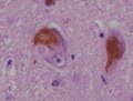 Lewy Koerperchen.JPG 331 × 251; 9 KB
 LIBRO TEXTO IPP HISTOLOGIA.pdf 1,275 × 1,650, 81 pages; 1.35 MB
 Lymph -.jpg 3,115 × 2,114; 1.15 MB
 Major Modes and Cellular Mechanisms of Secretion.png 1,501 × 517; 697 KB
 Mango Bark.jpg 3,072 × 2,304; 1.43 MB
 Medical Students.jpg 1,600 × 1,200; 288 KB
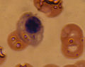
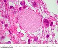
 Megakaryocyte.jpg 1,434 × 1,002; 1.4 MB
 Mesoderm hr.png 384 × 182; 59 KB
 Mesoderm-ar.png 384 × 184; 71 KB
 Mesoderm.png 384 × 182; 95 KB
 Metabolismo hueso.png 468 × 360; 24 KB
.JPG/120px-Metallose_(histologisch).JPG) Metallose (histologisch).JPG 2,046 × 1,364; 603 KB
Metallose (histologisch).JPG 2,046 × 1,364; 603 KB
 Metarubricyte.jpg 533 × 417; 143 KB
 Microscope slides prepared by a histologist in 1850. Wellcome M0011458.jpg 3,272 × 3,378; 3.18 MB
 Microscópio Óptico.png 647 × 595; 265 KB
 Mikroglej 1.jpg 480 × 357; 20 KB
 Mitoses anaplastic meningioma.jpg 2,080 × 1,542; 890 KB
 ModellEpithelformationJuxtaoralOrgan.jpg 425 × 93; 9 KB
 ModellJuxtaoralOrgan.jpg 354 × 345; 15 KB
 ModellrekonstruktionBeziehungNBuccalisJuxtaoralOrgan.jpg 354 × 329; 15 KB
_Cross-section.jpg/120px-Nasal_mucosa_(26_2_17)_Cross-section.jpg) Nasal mucosa (26 2 17) Cross-section.jpg 2,773 × 1,775; 1,021 KB
Nasal mucosa (26 2 17) Cross-section.jpg 2,773 × 1,775; 1,021 KB
.jpg/120px-Nasal_mucosa_-_human_(254_09).jpg) Nasal mucosa - human (254 09).jpg 3,750 × 2,400; 1.45 MB
Nasal mucosa - human (254 09).jpg 3,750 × 2,400; 1.45 MB
 Nerve cells. Wellcome M0016972.jpg 2,802 × 3,836; 4.02 MB
 Neuroglia de la region central gris Wellcome L0040801.jpg 2,666 × 3,423; 3.33 MB
 Neuronas multipolares en cerebro de ratón.jpg 3,840 × 3,072; 5.01 MB
 NeuroneChauveau1890MeyCh.jpg 960 × 1,576; 263 KB
 NguyenHue.jpg 1,000 × 767; 492 KB
 Nichtlaktierende Mama Elastika-Faerbung.gif 172 × 140; 25 KB
 Nueral tissue 200x magnification.jpg 2,619 × 2,684; 1,019 KB
.png/120px-OCT_embedding_(Optimal_Cutting_Temperature_compound).png) OCT embedding (Optimal Cutting Temperature compound).png 4,752 × 3,168; 11.47 MB
OCT embedding (Optimal Cutting Temperature compound).png 4,752 × 3,168; 11.47 MB
 Oesophageal layers.png 720 × 326; 151 KB
 Organe en bourrelet de Meta bourneti.jpg 1,196 × 858; 122 KB
 Organe gonoporal de Chiracanthium.jpg 1,481 × 1,127; 174 KB
 Organelles of the Secretory Pathway.png 1,035 × 1,126; 1.27 MB
 Organes gonoporaux d' Argyronète.jpg 2,633 × 2,214; 400 KB
 Osso - esponjoso e compacto.gif 636 × 336; 31 KB
 Osso longo regs.jpg 612 × 580; 80 KB
 Parietal cell miguelferig.png 1,379 × 1,194; 129 KB
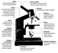
 Partes de un microscopio óptico.png 647 × 595; 160 KB
 Periferal nerve myelination.jpg 822 × 232; 120 KB
 Philaeus chrysops, région épigastrique.jpg 3,976 × 2,670; 691 KB
 Piel de humano.jpg 3,840 × 3,072; 6.84 MB
 Plasmazelle 2.jpg 133 × 153; 11 KB
 Pomescleroid40x1.jpg 1,024 × 768; 202 KB
 Psuedostratified epithelium animated.gif 373 × 394; 375 KB
 Queratinocitos del estrato espinoso de piel gruesa de mamífero.jpg 2,560 × 2,048; 2.19 MB
 Raji cell lines.jpg 1,077 × 610; 138 KB
 RatBarrelFieldCOstain.jpg 2,048 × 1,536; 646 KB
 Rectus abdominis muscle 2.jpg 960 × 720; 86 KB
 Respiratory Tract Cells.png 964 × 918; 1.04 MB
 Respiratory Tract Histological Differences.png 1,500 × 964; 807 KB
 Rubricyte.jpg 960 × 756; 387 KB
 Région épigastrique Pholcus.jpg 3,468 × 2,562; 586 KB
 Rôsolovité väzivo, Wharton's jelly - histológia, histology.jpg 2,542 × 2,304; 2.29 MB
 Saco polinico.JPG 1,173 × 798; 155 KB
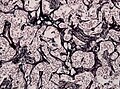
 Scar collagen.jpg 9,676 × 6,437; 4.46 MB
 Sclerochronologie-Nl.png 1,282 × 1,978; 58 KB
 Sehnenscheide.jpg 213 × 189; 15 KB
 Serous organ invagination.gif 443 × 192; 6 KB
 Sitios matricripticos.jpeg 1,800 × 1,236; 260 KB
 Slide rack in haematoxylin.jpg 1,731 × 1,853; 1.48 MB
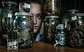
 Slide storage in histology.jpg 2,000 × 2,500; 2.57 MB
 Slide under a microscope.jpg 2,568 × 3,015; 1.7 MB
 Smith.PNG 1,500 × 928; 735 KB
 Stained microscope slide.jpg 2,299 × 1,731; 575 KB
 Stratified cuboidal epithelium animated.gif 665 × 535; 976 KB
 Synovium.png 618 × 88; 7 KB
 T. Schwann, Mikroskopische Untersuchungern u Wellcome L0032288.jpg 1,732 × 1,296; 951 KB
 T. Schwann, Mikroskopische Untersuchungern u Wellcome L0032289.jpg 1,704 × 1,264; 926 KB
 T. Schwann, Mikroskopische Untersuchungern u Wellcome L0032290.jpg 5,062 × 3,796; 1.74 MB
 Terminaciones nerviosas en la piel y pelos del raton Wellcome L0040802.jpg 3,254 × 2,672; 1.7 MB
 The cell structure of tissues, etc. Wellcome M0011241.jpg 3,862 × 2,961; 2.71 MB
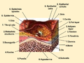
.jpg/112px-Thick_Skin_Histological_Section_(epidermis%2C_dermis_and_hypodermis).jpg) Thick Skin Histological Section (epidermis, dermis and hypodermis).jpg 1,536 × 1,644; 576 KB
Thick Skin Histological Section (epidermis, dermis and hypodermis).jpg 1,536 × 1,644; 576 KB
 Tissue MicroArray Slide.jpg 647 × 480; 26 KB
 TLP Verband 2.jpg 674 × 452; 104 KB
 TLP-Verband 1.jpg 684 × 451; 86 KB
 Topping-type microtome, Europe, 1840-1850 Wellcome L0058210.jpg 2,676 × 3,921; 1.21 MB
 Transfection monocyte.jpg 1,500 × 1,000; 256 KB
 Transitional epithelium animated.gif 561 × 549; 646 KB
 Traqueia – HE – 400x.png 1,520 × 1,137; 3.51 MB
 Tumor of follicular infundibulum.jpg 2,448 × 1,920; 1.05 MB
 Turbellaria.jpg 6,242 × 3,061; 1.7 MB
 Uroctea, glande segmentaire du rostre.jpg 2,635 × 3,434; 850 KB
 Uroctea, organe du pharynx et glande segmentaire rostrale.jpg 1,469 × 1,928; 362 KB
 Uroctea, organe pharyngien, détail.jpg 1,466 × 1,916; 350 KB
 Uroctea, organe pharyngien.jpg 1,451 × 1,936; 297 KB
 Uroctée pharyngien 1.jpg 3,473 × 2,393; 534 KB
 Uroctée pharyngien 2.jpg 3,959 × 2,489; 642 KB
 Valentin knife, London, England, 1850-1870 Wellcome L0057585.jpg 4,256 × 2,832; 1.35 MB
 Vas deferens Histology.jpg 2,048 × 1,536; 337 KB
 Vas deferens.jpg 2,816 × 2,112; 756 KB
 Vibratome.jpg 1,536 × 2,048; 1.03 MB
 Voronoi on neurons.jpg 1,278 × 641; 449 KB
 Wallerian degeneration of a nerve. Wellcome L0002019.jpg 1,228 × 1,520; 474 KB
 Wavy collagen.jpg 9,672 × 6,456; 2.82 MB
 Xantoastrocitoma Pleomórfico de células gigantes.jpg 640 × 549; 107 KB
 Young scientist PhD.jpg 4,152 × 2,598; 14.97 MB
 Zapfenverschaltung Netzhaut.jpg 601 × 351; 23 KB
 Zellballen paraganglioma.jpg 2,080 × 1,542; 1,003 KB
 Žaizdų gylis odoje.png 960 × 720; 509 KB
.jpg/120px-Дикроцелий_Ланцетовидный_сосальщик_(13).jpg) Дикроцелий Ланцетовидный сосальщик (13).jpg 4,272 × 2,848; 2.31 MB
Дикроцелий Ланцетовидный сосальщик (13).jpg 4,272 × 2,848; 2.31 MB


_-_IHM-0651.jpg)


_-_copia.jpg)


.jpg)
.jpg)
.jpg)

.jpg)


.jpg)


.JPG)


_Cross-section.jpg)
.jpg)


.png)


.jpg)


.jpg)


.jpg)
.jpg)
.jpg)