Category:Molecular biology
Jump to navigation
Jump to search

branch of biology that deals with the molecular basis of biological activity  | |||
| Upload media | |||
| Instance of | science | ||
|---|---|---|---|
| Subclass of | biology | ||
| Part of | biology | ||
| Has part |
| ||
| |||
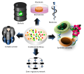
English: Molecular biology is the study of biology at a molecular level. The field overlaps with other areas of biology, particularly genetics and biochemistry. Molecular biology chiefly concerns itself with understanding the interactions between the various systems of a cell, including the interrelationship of DNA, RNA and protein synthesis and learning how these interactions are regulated.
Subcategories
This category has the following 79 subcategories, out of 79 total.
*
B
C
D
E
F
G
H
I
K
M
N
O
P
S
T
W
Pages in category "Molecular biology"
The following 2 pages are in this category, out of 2 total.
Media in category "Molecular biology"
The following 200 files are in this category, out of 639 total.
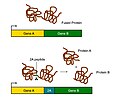
(previous page) (next page)2A_peptide_Working_Mechanism.jpg/120px-(zh)2A_peptide_Working_Mechanism.jpg) (zh)2A peptide Working Mechanism.jpg 593 × 493; 125 KB
(zh)2A peptide Working Mechanism.jpg 593 × 493; 125 KB
 031-P53-1olg 1tup 1ycq.gif 481 × 553; 81 KB
 1-3 BPG in active site.png 341 × 415; 38 KB
 1M7 RNA adduct.png 800 × 365; 12 KB
 24s-ohc.jpg 626 × 429; 20 KB
 2A peptide Working Mechanism.jpg 593 × 493; 129 KB
 2D gel images dual channel original.PNG 374 × 265; 60 KB
 2D gel images dual channel warped.PNG 374 × 265; 57 KB
 3D pyruvate dehydrogenase complex.jpg 4,800 × 5,400; 5.18 MB
 3primeR2.jpg 1,523 × 1,492; 67 KB
 4nqo1.gif 521 × 119; 6 KB
 4nqo2.png 784 × 194; 60 KB
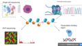
 AAV Gene Therapy.jpg 495 × 371; 137 KB
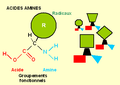 Acides aminés.PNG 370 × 263; 8 KB
 Acquaporina subunità porocanale.png 986 × 492; 27 KB
 Actin related proteins.jpg 1,200 × 1,200; 192 KB
 Active Site 1.png 491 × 366; 72 KB
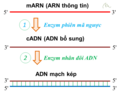 ADN bổ sung Complementary DNA.png 684 × 540; 23 KB
 ADN clonage.png 516 × 98; 13 KB
 ADN extraction.png 613 × 174; 23 KB
 ADN hybridation.png 611 × 127; 16 KB
 ADN purification.png 613 × 127; 19 KB
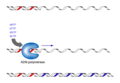 ADNpol.png 375 × 260; 16 KB
 Affinity Chromatography.jpg 580 × 800; 161 KB
 Affymetrix 5.0 microarray.jpg 1,566 × 2,349; 1.73 MB
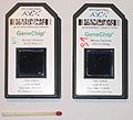
 Affymetrix GeneChip.jpg 750 × 600; 312 KB
 Affymetrix-microarray.jpg 665 × 603; 130 KB
 Ahelix-FMO-Facio.jpg 1,073 × 821; 105 KB
 Alphahelix.png 651 × 757; 141 KB
 Amine reaction.png 440 × 160; 3 KB
 Amino acid substitution pattern in epPCR and SeSaM.tif 1,246 × 528; 440 KB
 Annotated Theoretical Model of Bound Tetrameric Lac Repressor.png 1,162 × 1,010; 679 KB
 Annular Gap Junction Vesicle.jpg 205 × 153; 61 KB
 Apobgene.PNG 605 × 534; 36 KB
 Apotosis.jpg 1,302 × 520; 157 KB
 Application of MNAse.png 959 × 540; 194 KB
 Applications of combinatorial gene circuit optimization strategies.svg 512 × 451; 217 KB
 Arnmensajero1.png 283 × 212; 28 KB
 ASO dot blot inverso.png 1,310 × 786; 42 KB
 ASO dot blot.png 1,304 × 864; 38 KB
 Babraham-towards gate.JPG 2,560 × 1,920; 355 KB
 Babrahamback.JPG 1,920 × 2,560; 428 KB
 Babrahamtowardscience.JPG 2,560 × 1,920; 389 KB
 BAC work flow ESP.jpg 1,234 × 1,113; 75 KB
 BACs cloning vectors Chem114A.jpg 896 × 921; 70 KB
 Balizas moleculares.png 960 × 720; 11 KB
 Bcrablinter.jpg 349 × 337; 10 KB
 Beta-catenin-in-cancer.png 776 × 744; 43 KB
 Beta-catenin-moonlighting.png 562 × 746; 69 KB
 Beta-ketothiolase.png 814 × 880; 62 KB
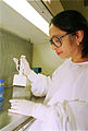
 Bilal Djeghout Laboratory.jpg 1,966 × 2,048; 361 KB
 Binding.png 888 × 572; 22 KB
 Biologia molecolare - Biochimica - Genetica.jpg 1,065 × 871; 74 KB
_Front_view.jpg/120px-Biological_Safety_Cabinet_(Class_II%2C_Type_A2)_Front_view.jpg) Biological Safety Cabinet (Class II, Type A2) Front view.jpg 1,469 × 1,102; 420 KB
Biological Safety Cabinet (Class II, Type A2) Front view.jpg 1,469 × 1,102; 420 KB
_Side_view.jpg/120px-Biological_Safety_Cabinet_(Class_II%2C_Type_A2)_Side_view.jpg) Biological Safety Cabinet (Class II, Type A2) Side view.jpg 1,469 × 1,102; 503 KB
Biological Safety Cabinet (Class II, Type A2) Side view.jpg 1,469 × 1,102; 503 KB
_Telstar_front_view.jpg/120px-Biological_Safety_Cabinet_(Class_II%2C_Type_A2)_Telstar_front_view.jpg) Biological Safety Cabinet (Class II, Type A2) Telstar front view.jpg 2,048 × 1,536; 731 KB
Biological Safety Cabinet (Class II, Type A2) Telstar front view.jpg 2,048 × 1,536; 731 KB
 Biosensors used for screening combinatorial DNA libraries.svg 512 × 256; 122 KB
.png/120px-BLESS_workflow_(update_v2).png) BLESS workflow (update v2).png 1,677 × 764; 214 KB
BLESS workflow (update v2).png 1,677 × 764; 214 KB
 Bloodgroup-Blutgruppen-Glyk.jpg 1,024 × 768; 66 KB
 Blotautoradiogram.png 124 × 105; 7 KB
 Brefeldin A Inhibition of Intracellular Vesicle Transport.png 591 × 271; 23 KB
.png/69px-Burtyka_alphabet_of_Genetic_language_(BaGl).png) Burtyka alphabet of Genetic language (BaGl).png 801 × 1,384; 230 KB
Burtyka alphabet of Genetic language (BaGl).png 801 × 1,384; 230 KB
 Burtykon - the letter of the Genetic alphabet.png 1,243 × 524; 34 KB
 Caged ATP and cAMP.jpg 937 × 422; 36 KB
.jpg/120px-Caged_ATP_cAMP_(+Br).jpg) Caged ATP cAMP (+Br).jpg 378 × 221; 9 KB
Caged ATP cAMP (+Br).jpg 378 × 221; 9 KB
 Capillarywall.png 436 × 217; 6 KB
 Cascada JNK.jpg 2,001 × 1,501; 194 KB
 Cassettes.png 960 × 720; 42 KB
 CCT gamma apical.png 1,060 × 709; 360 KB
 CDNA-library-amplification.png 2,955 × 1,419; 159 KB
 CE Basic.jpg 529 × 320; 19 KB
 Central Dogma Model.png 800 × 557; 53 KB
 CGDB CGDef.png 775 × 200; 30 KB
 CGDef 1.png 693 × 179; 28 KB
 ChIPPET3.JPG 720 × 540; 24 KB
 ChIPPET4.JPG 720 × 540; 16 KB
 ChIPPET5.JPG 720 × 540; 19 KB
 ChIPPET6.JPG 720 × 540; 13 KB
 ChIPPET7.JPG 720 × 540; 13 KB
 ChIPPET8.JPG 720 × 540; 32 KB
 ChIPPET9.JPG 720 × 540; 26 KB
 Chitinase4TC.jpg 420 × 260; 55 KB
 Choosing a cloning method.png 2,625 × 1,425; 144 KB
 Chromatogramme.jpg 583 × 768; 51 KB
 Chélation.jpg 455 × 218; 19 KB
 Codones-ARN.png 225 × 382; 18 KB
 ColonneADN.JPG 153 × 359; 4 KB
 ColonneElution.JPG 153 × 359; 6 KB
 ConceptLarge.jpg 97 × 380; 28 KB
 Conceptual Translation 1.jpg 642 × 153; 20 KB
 Conceptual Translation.jpg 683 × 847; 132 KB
 Condensation3.png 2,852 × 791; 279 KB
 ConservedDomains.png 1,098 × 674; 392 KB
 Consumo excesivo de alimentos altos en Triacilgliceroles.jpg 897 × 701; 158 KB
 Conventional RNAi.png 3,562 × 2,160; 683 KB
 Coomassie3.jpg 840 × 1,573; 107 KB
 COREcassette.PNG 960 × 720; 43 KB
 Courtney 2008.jpg 786 × 685; 52 KB
 CreLoxP.jpg 320 × 316; 13 KB
 Crystal structure of the zinc finger type C2H2 from A. thaliana.png 830 × 559; 33 KB
 Crystal-by-example-illustrating-how-a-helix-might-be-formed.jpg 600 × 514; 82 KB
 CSIRO ScienceImage 1579 Cane toad.jpg 2,657 × 1,767; 4.32 MB
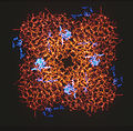
 CSIRO ScienceImage 1788 Molecular model of a protein.jpg 2,657 × 1,774; 3.12 MB
 CSIRO ScienceImage 1789 Protein.jpg 2,694 × 2,657; 5.12 MB
 CSIRO ScienceImage 1802 Protein model.jpg 1,834 × 2,657; 4.04 MB
 CSIRO ScienceImage 1900 Fowl Adenovirus.jpg 945 × 743; 563 KB
 CSIRO ScienceImage 1916 Milking a Transgenic Sheep.jpg 2,660 × 2,657; 4.4 MB
 CSIRO ScienceImage 1953 Difference Between Transgenic Sheep and Normal.jpg 2,682 × 2,657; 5.08 MB
 CSIRO ScienceImage 2082 Cell Culture.jpg 2,657 × 1,976; 5.15 MB
 CSIRO ScienceImage 2084 Helen Newton Turner 19081995.jpg 1,772 × 1,337; 911 KB
 CSIRO ScienceImage 2112 Transgenic Sheep in a Field.jpg 2,706 × 2,657; 5.68 MB
 CSIRO ScienceImage 2294 A normal sized control sheep left and transgenic sheep right.jpg 2,660 × 2,657; 5.86 MB
 CSIRO ScienceImage 2465 Sequencing DNA.jpg 1,179 × 1,772; 1.77 MB
 CSIRO ScienceImage 2589 Shape of Protein Backbone.jpg 2,657 × 1,760; 4.95 MB
 CSIRO ScienceImage 2738 First Three Domains of Insulin.jpg 1,712 × 2,657; 4.25 MB
 CSIRO ScienceImage 2993 Old Amino Acid Sequencer.jpg 1,813 × 2,657; 3.3 MB
 CSIRO ScienceImage 7305 A group of transgenic sheep.jpg 2,707 × 2,657; 5.24 MB
 CSIRO ScienceImage 86 Developing an Enzyme Linked Immunosorbent Assay.jpg 1,793 × 2,657; 4.98 MB
 Cytosine becomes thymine ar.png 1,280 × 582; 87 KB
 Cytosine becomes thymine.png 5,040 × 2,293; 44.16 MB
 DdATP.png 219 × 216; 6 KB
 Deconvoluted ESMS.jpg 1,648 × 1,123; 71 KB
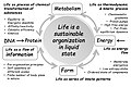 Definition of cellular life NB.jpg 720 × 479; 188 KB
 Dien bien nhanh.JPG 478 × 709; 64 KB
 Digestión, absorción y metabolismo de ácidos grasos.jpg 1,018 × 814; 215 KB
 DLD1 HUMAN Gel.gif 660 × 691; 336 KB
 DNA methyltransferases and EC system.png 2,835 × 1,595; 796 KB
 DNA microarray.jpg 280 × 285; 40 KB
 DNA molecular structure, showing individual nucleotides and bonds.jpg 693 × 984; 110 KB
 DNA Nucleotides.jpg 817 × 408; 185 KB
 DNA sequence, Nucleotide sequence.gif 972 × 2,072; 1.51 MB
 DNA transposition.gif 365 × 339; 51 KB
 DNA-Leiter.jpg 404 × 802; 55 KB
 Dna-microarray.jpg 422 × 464; 42 KB
 DNA-RNA D-loops, greatly exaggerated.jpg 2,562 × 2,731; 243 KB
 DNaseI footprint.png 162 × 626; 79 KB
 Donning.jpg 1,102 × 1,469; 440 KB
 Draft figures 1 - 4 from second Watson and Crick paper Wellcome L0032989.jpg 3,256 × 4,072; 1.41 MB
 Droga alternatywna.png 759 × 350; 66 KB
 Droga klasyczna.png 725 × 405; 74 KB
 Dsx splicing.jpg 876 × 499; 83 KB
 Duplex sequencing library preparation procedure.png 1,053 × 1,559; 111 KB
 Duplex sequencing overview alphabeta fix.svg 1,417 × 1,536; 9.57 MB
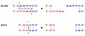
 Early genetic code.jpg 551 × 552; 106 KB
 Early genetic code.svg 512 × 512; 30 KB
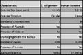 Ecoli human compare.jpg 675 × 450; 51 KB
 Editing.PNG 601 × 337; 55 KB
 Electrofiltration filterplate.jpg 322 × 269; 11 KB
_using_magnetic_swipe_card.jpg/120px-Electronic_access_control_(BSL3_Lab)_using_magnetic_swipe_card.jpg) Electronic access control (BSL3 Lab) using magnetic swipe card.jpg 2,048 × 1,536; 702 KB
Electronic access control (BSL3 Lab) using magnetic swipe card.jpg 2,048 × 1,536; 702 KB
_using_personal_identification_number_(PIN).jpg/120px-Electronic_access_control_(BSL3_Lab)_using_personal_identification_number_(PIN).jpg) Electronic access control (BSL3 Lab) using personal identification number (PIN).jpg 2,048 × 1,536; 732 KB
Electronic access control (BSL3 Lab) using personal identification number (PIN).jpg 2,048 × 1,536; 732 KB
.jpg/120px-Electronic_access_control_(BSL3_Lab).jpg) Electronic access control (BSL3 Lab).jpg 2,048 × 1,536; 725 KB
Electronic access control (BSL3 Lab).jpg 2,048 × 1,536; 725 KB
 Electronic-adressing.jpg 421 × 228; 22 KB
 Electroosmoticflow.png 406 × 91; 2 KB
 Electrophoretic separation of circular DNA strands.gif 88 × 400; 8 KB
 Electroporation Cuvettes.jpg 1,024 × 768; 138 KB
 Electroporation Diagram.png 832 × 512; 17 KB
 Elongation der Translation.png 1,751 × 584; 153 KB
 Endonucleolyticcleavage.jpg 582 × 695; 28 KB
 Epissage.png 565 × 222; 10 KB
 Erkennungssequenzen von Restriktionsenzymen.jpg 764 × 331; 22 KB
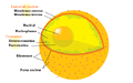 Esquema de nucli cel·lular.PNG 552 × 346; 45 KB
 EsquemaBiologiaMolecular.png 576 × 354; 599 KB
 Esructura 3D del ácido perfluorooctanoico.jpg 317 × 169; 43 KB
 Estructura 3D del sulfonato de perfluorooctano.jpg 342 × 161; 16 KB
 Estructura ORC.png 399 × 216; 21 KB
 EvolutionOfDuplicateGenes.png 1,326 × 677; 28 KB
 Exon intron.png 517 × 93; 5 KB
 Experimento-pulso-caza.jpg 500 × 272; 39 KB
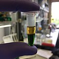 Extraction DNA.jpg 2,448 × 2,448; 903 KB
 Extrapolation based Molecular Systems biology GRana 09.jpg 960 × 720; 73 KB
 Extrapolation based Molecular Systems biology Rana 09.jpg 960 × 720; 73 KB
 Extrapolation Based Molecular Systems Biology Rana CWRU.tiff 960 × 720; 589 KB
_collected_in_Sweden.jpg/120px-Faeces_of_wolf_(Canis_lupus)_collected_in_Sweden.jpg) Faeces of wolf (Canis lupus) collected in Sweden.jpg 2,272 × 1,704; 776 KB
Faeces of wolf (Canis lupus) collected in Sweden.jpg 2,272 × 1,704; 776 KB
 Faire scheme.png 3,000 × 2,300; 1.78 MB
 Fedg.png 1,086 × 817; 363 KB
 Fig2.Recombination patterns.png 960 × 720; 12 KB
 Finite Element Model.jpg 793 × 634; 41 KB
 Finite model.jpg 1,050 × 734; 53 KB
.jpg/120px-Fish_Egg_Diagram_(1).jpg) Fish Egg Diagram (1).jpg 960 × 720; 38 KB
Fish Egg Diagram (1).jpg 960 × 720; 38 KB
 Fish Egg.jpg 960 × 720; 37 KB
 FISHchip.jpg 1,392 × 1,160; 771 KB
 FlAsh Protein Modification.png 2,161 × 676; 110 KB
 Flusso informazione genetica diagramma.jpg 854 × 234; 37 KB
 Footprinting DNA.png 745 × 700; 34 KB
 Fret.jpg 640 × 439; 20 KB
 Fret.svg 640 × 439; 21 KB
 Functional Cloning.png 5,500 × 800; 611 KB
 G-quadruplex gene regulation model.jpg 647 × 335; 33 KB
 G-quadruplex.gif 500 × 300; 11 KB
 Gel elecro.JPG 2,800 × 2,128; 2.78 MB
 Gelelektroforese.jpg 535 × 314; 14 KB
 Gene expression control.png 800 × 581; 90 KB
.svg/120px-Gene2-plain_(zh-cn).svg.png) Gene2-plain (zh-cn).svg 1,395 × 1,182; 30 KB
Gene2-plain (zh-cn).svg 1,395 × 1,182; 30 KB
 Gene2-plain-norsk.svg 1,395 × 1,182; 27 KB
 Gene2-plain.svg 1,395 × 1,182; 29 KB
2A_peptide_Working_Mechanism.jpg)


_Front_view.jpg)
_Side_view.jpg)
_Telstar_front_view.jpg)

.png)


.png)


.jpg)


_using_magnetic_swipe_card.jpg)
_using_personal_identification_number_(PIN).jpg)
.jpg)


_collected_in_Sweden.jpg)


.jpg)


.svg)

